When examining a medical endoscope, we might first notice a small lens or a round glass surface. However, the technical term for this area is distal tip; the distal tip houses a highly complex miniature lens assembly. It is the core part of the entire endoscope, responsible not only for capturing and transmitting images but also for providing illumination, accommodating channels for surgical instruments, and ensuring clinical safety and precision. Different types of distal tips are configured to suit the anatomical structures of specific organs and the needs of surgical observation.
The design of the distal assembly directly determines image quality, operational precision, and surgical outcomes. A precisely engineered distal tip allows surgeons to identify tissue details during minimally invasive procedures clearly.
Types of Endoscopes – Rigid vs. Flexible
Rigid Endoscopes
Rigid endoscopes are commonly used in procedures such as arthroscopy and cystoscopy. Their rigid tube structure allows for the integration of high-resolution lenses and large-diameter optical components, providing clearer and more stable images. The optical lens assembly is typically small and robust, with a fixed angle of view, and is suitable for procedures that require precise positioning and high-resolution imaging.
Flexible Endoscopes
Flexible endoscopes are widely used in gastroenterology, nasopharyngoscopy, and bronchoscopy, where navigation through curved or complex pathways is required. The optical lens assembly typically contains 7–8 lenses, often combined with miniature CMOS sensors or fiber optic image bundles to ensure stable image transmission even when the scope is bent. While offering superior maneuverability, the optical components are extremely small, requiring high precision and miniaturization without compromising clarity.
Hybrid Designs
Some minimally invasive surgical endoscopes combine rigid and flexible structures, featuring detachable distal tips or curved rigid tube designs. These hybrid designs leverage the advantages of both rigid and flexible scopes, ensuring high-definition image quality while providing flexibility for complex examinations and surgical procedures.
Specific Clinical Applications
Nasal Endoscopes
Nasal endoscopes are slender and compact. Their optical lens assembly emphasizes high resolution and a wide field of view within narrow spaces, allowing for the detailed observation of fine structures and lesions.

Gastrointestinal Endoscopes
The distal tip of a gastrointestinal endoscope has a larger diameter to accommodate an instrument channel. The assembly typically integrates a light source, camera, air/water nozzle, and suction channel, supporting complex diagnostic and therapeutic procedures.

Bronchoscopes and Ureteroscopes
These endoscopes feature a flexible distal tip capable of navigating complex pathways. The assemblies are multifunctional, supporting both imaging and instrument channels, as well as air/water functions, meeting the requirements of minimally invasive surgery and diagnostic procedures.

The Protective Window
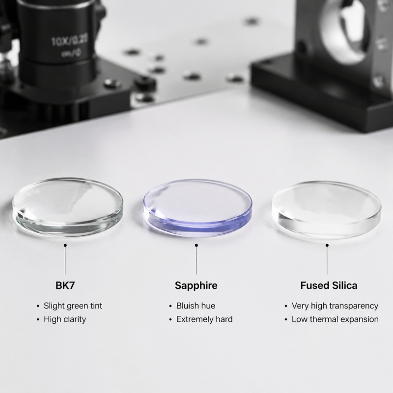
The protective optical window at the distal tip is typically made of sapphire, optical glass, or medical-grade polymer. It must resist scratches, fog, and contamination, while withstanding high-temperature and chemical sterilization. Modern designs may incorporate multiple surface coatings depending on specific requirements—such as anti-reflective coatings to improve light transmittance, oleophobic coatings to reduce fingerprints and bodily fluid adhesion, and other treatments to enhance image clarity. Whether these coatings are applied depends on the endoscope type, clinical application, and imaging needs. The thickness and curvature of the window may also vary to suit specific anatomical sites and imaging requirements.

Optical Imaging System
The optical lens assembly at the distal tip typically consists of multiple miniature lenses, optimized according to design requirements. Common lens types include plano-convex, plano-concave, biconvex, biconcave, meniscus (convex-concave), as well as cemented lenses and cemented prisms used for aberration correction or compact optical layouts. In some designs, a sapphire protective element is integrated at the front to enhance wear resistance and environmental durability.

Aspheric lenses may also be incorporated to reduce spherical aberration and distortion in a limited space, improving image clarity. However, the use of aspheric elements depends on size constraints, cost, and performance requirements; not all endoscopes include them.
Different endoscope types have distinct optical design priorities. Nasal endoscopes focus on high-resolution close-range imaging for fine tissue structures, while gastrointestinal endoscopes emphasize a wide field of view and uniform depth of field for broader observation. Overall, the configuration of the lens assembly directly affects resolution, distortion control, and imaging uniformity, which in turn influences clinical diagnostic accuracy and operational stability.
Illumination Function
The distal tip provides both image capture and stable illumination. Flexible endoscopes typically use fiber optic light guides to transmit light from an external cold light source to the examination area, minimizing thermal effects on tissues. Some modern systems integrate miniature LED light sources directly into the distal tip, enabling more compact designs and providing relatively uniform, controllable illumination.
Light source selection is constrained by distal tip size, structural space, and heat dissipation. Rigid endoscopes, with stable and spacious distal tip, can accommodate higher-power external light sources, while flexible endoscopes rely on fiber optics or miniaturized LEDs to balance illumination, reliability, and flexibility.
Image Transmission
Traditional flexible endoscopes transmit images from the distal tip to external displays via fiber optic bundles, where image fidelity is primarily governed by fiber count, packing density, and transmission attenuation. In contrast, digital endoscopes integrate miniature CMOS or CCD sensors directly into the distal tip. By converting optical signals to electrical data at the source, these systems enable significantly higher resolution and more stable, real-time imaging.
Rigid endoscopes, leveraging a fixed optical path and stable architecture, can accommodate larger-aperture lenses and premium optical components. This results in superior clarity and exceptional detail reproduction. However, their inherent rigidity limits maneuverability when navigating curved or complex anatomical pathways.

Additional Functional Channels
The distal tip typically integrates multiple functional channels, including instrument channels, air/water jet channels, and suction channels. Channel configurations vary according to clinical requirements, resulting in significant differences among endoscope types.
For example, gastrointestinal endoscopes feature relatively large channels for instruments, combined with air/water jet and suction capabilities to support cleaning, dilation, and auxiliary operations. Nasal endoscopes and bronchoscopes emphasize compactness and flexibility, optimizing channel size and quantity to navigate narrow or curved anatomical structures while minimizing interference with imaging.
Future Clinical Trends
Endoscope distal tips are evolving toward miniaturization, integration, and intelligence. AI-assisted image processing may enhance lesion detection, while 3D and multispectral imaging technologies provide richer tissue information, offering more dimensions for clinical assessment.
Single-use endoscopes are gradually emerging to reduce cross-infection risks and simplify instrument maintenance. Although the design priorities vary for nasal, gastrointestinal, and rigid endoscopes, the overall trend is toward higher performance, greater integration, and enhanced safety.
Conclusion
The distal tip of a medical endoscope is not merely a lens or protective window; it is a precision-engineered assembly that integrates optics, electronics, mechanics, and fluid systems. Its design directly affects image clarity, operational precision, and clinical outcomes. These small yet complex structures make the medical endoscope an indispensable tool in modern minimally invasive medicine.